FIG. 6.
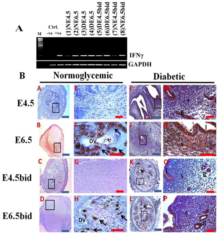
Detection of changes in uterine IFNγ gene expression by semiquantitative PCR (A) and in histological sections (B) in the normally mated and ConA-induced pseudopregnant cNOD and dNOD mice during peri and postimplantation. A) IFNγ mRNA was expressed in high levels in the uteri of dNOD mice throughout early pregnancy and/or pseudopregnancy days 4.5 and 6.5, respectively (A, compare lanes 3–6 with lanes 1–2 and 7–8, respectively). Transcripts from lipopolysaccharides-stimulated mouse peritoneal cells tested for INFγ mRNA were loaded to the positive (+ve) control lane, and IFNγ complementary sense sequence “gtctggcctgctgttaaagc” was added to the PCR reaction to generate a negative (-ve) control. B) Time-course IHC detection of uterine IFNγ in histological sections of cNOD (A–H) and dNOD (I–P) mice at the peri- and postimplantation, and in the ConA-induced pseudopregnancy. E–H and M–P show at higher magnifications of the corresponding inset images in A–D and I–L, respectively, featuring IFNγ immunolocalization in uNK cells on E6.5 (F, arrows) and E6.5bid (H, arrows). A diffuse epithelial and stromal immunolabeling was detected in the uteri of dNOD mice on E4.5, E6.5, E4.5bid and E6.5bid (M–P), respectively; n = 3 mice per group per 3 sets of independent PCR experiments with similar outcome. DV, decidual vessels; * indicates uterine lumen; l, luminal epithelium; s, stroma; g, metrial glands. Blue bar = 100 μm; red bar = 50 μm

