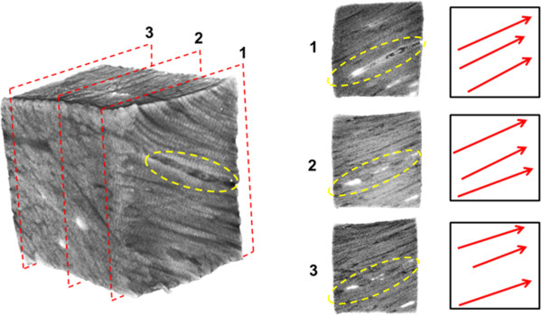
Figure 2.

A meniscal sample taken from the outer one-third of the main body of the meniscus. The planes identified by the red dashed lines are shown in the panels 1-3 to the right. Predominant fascicle directions are illustrated by the red arrows. All planes showed similar fascicle orientations. The collagen sparse void space containing blood vessels are indicated by the yellow dashed ellipses.
