
Figure 4.
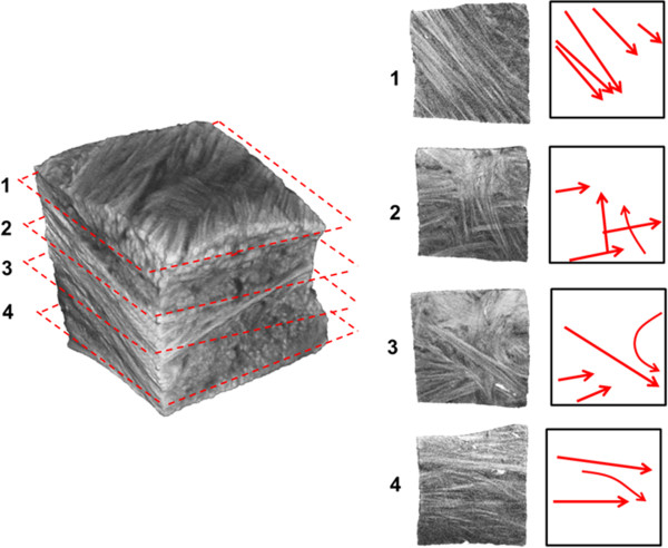
A meniscal sample taken from the inner one-third of the main body of the meniscus. Varying fascicle orientations can be observed in planes 1-4, moving in the superior to inferior direction. Red arrows to the right indicate the predominant fascicle directions in each breakout section image.