
Figure 3.
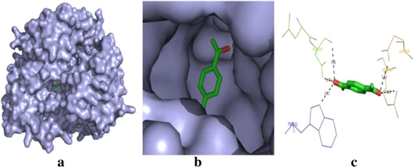
Molecular docking result of compound 1. (a) Docked poses of compound 1 in human phosphoinositide 3-kinase binding site. (b) A close-up view of the docked pose of compound 1; protein structure is shown in the surface model, and the ligand is shown in the stick model (color by atom). (c) H bond networks and bond distance are shown.