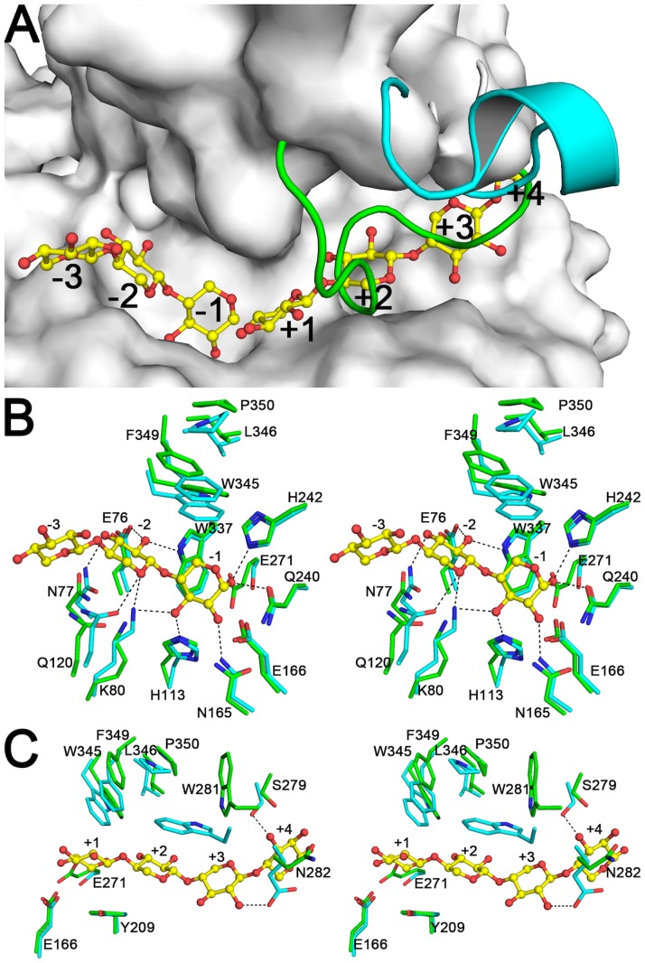
Figure 7. Substrate binding sites of SCXyl.
A) Molecular surface of chain A, with the Trp281-Arg291 loop from chain B and CmXyn10B shown as green and blue lines, respectively. B) Stereo view of the glycone region (from −3 to −1 subsites) of SCXyl (carbon atoms in green) and CmXyn10B (carbon atoms in blue). C) Stereo view of the aglycone region (from +1 to +4 subsites). The substrate found in the CmXyn10B-complex structure (PDB code: 1UQY) is shown in Figs. A, B and C, as ball-and-sticks. Residues numbering refers to the SCXyl enzyme.

