
Figure 1.
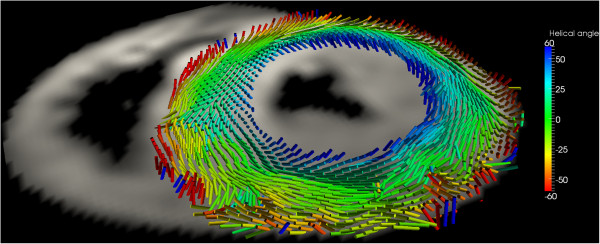
Diffusion tensor imaging in the basal short-axis plane in a patient with hypertrophic cardiomyopathy. The acquisition used a diffusion-weighted stimulated-echo single-shot echo-planar-imaging sequence during multiple breath holds. The tensor’s main eigenvector, which follows the orientation of the myocytes, is represented by cylindrical glyphs, colour coded according to the helix angle: blue right-handed, green circumferential, red left-handed (Image courtesy of Pedro Ferreira and Dudley Pennell).