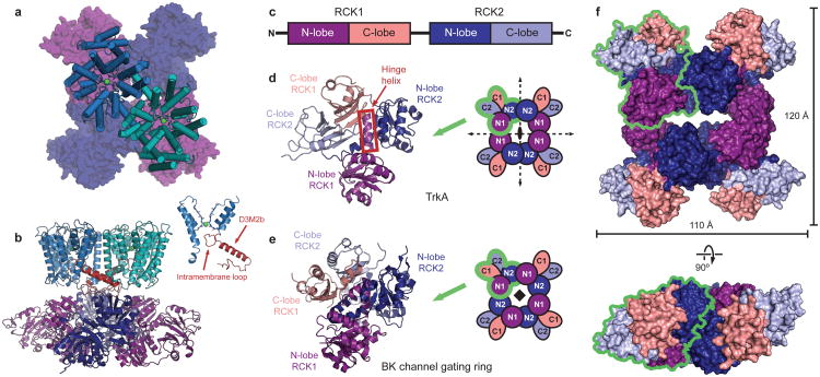
Figure 2. Structure of the TrkH-TrkA complex.
a.Structure of the complex between the TrkH dimer (light blue and teal) and the TrkA tetramer (dark blue and purple), viewed from the periplasmic side. The TrKA protomer is shown as a surface representation. b. The TrkH-TrkA complex viewed from within the plane of the membrane. Helix D3M2b in each TrkH subunit is highlighted in red. The inset on right shows homologous domains 1 and 3 of TrkH, with the intramembrane loop and helix D3M2b highlighted. c. Schematic showing the organization of RCK domains in a TrkA subunit. d-e. Cartoon representation of a TrkA protomer (d) or a protomer from the BK channel gating ring (e), colored by subdomain according to the color scheme in panel c. Schematics illustrating the organization of the tetrameric gating ring with the two-fold or four-fold symmetry axes marked are shown on the right. The green outlines delineate an individual subunit in each tetramer. f. Surface representation of the TrkA tetramer in the TrkH-TrkA complex, viewed from the periplasmic side (top) or parallel to the membrane (bottom). The green outline delineates an individual subunit.

