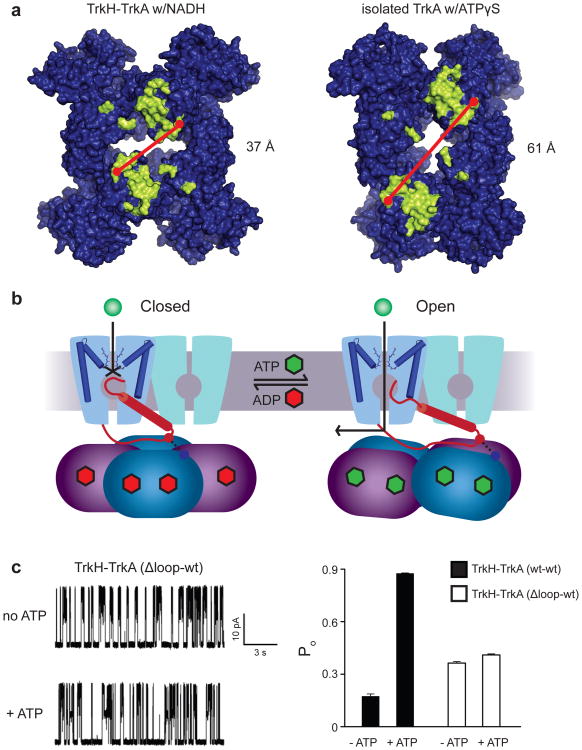
Figure 4. Proposed mechanism of regulation of TrkH gating by TrkA.
a. Surface representation of the TrkA tetramer from the TrkA-only (top) and TrkH-TrkA structures, viewed from the membrane-facing side. Residues forming the channel-gating ring interface, defined as residues with at least one atom within 4 Å of TrkH in the complex structure, are marked in yellow. b. Diagram illustrating a possible gating mechanism, with the closed channel shown on the left, and the open channel on the right. The D3M2b helix is represented as a red cylinder. For clarity, gating of only one TrkH protomer is shown. c. Left panel, single-channel currents through the TrkH-TrkA (Δ-loop-wt) complex before and after addition of ATP. The holding potential is +50 mV. Right panel, the Popen of TrkH-TrkA (wt-wt) and TrkH-TrkA (Δ-loop-wt) before and after addition of 5mM ATP. The error bars are s.e.m from 3 independent patches.

