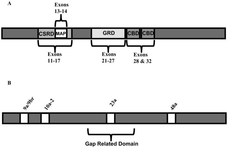
Figure 1.
Schematic representations of neurofibromin protein domains and alternative exons. (A) Neurofibromin protein with important domains and the exons which encode them highlighted. Protein domains are shown as grey or white boxes. The MAP domain is found within the CSRD. (B) Neurofibromin protein with naturally occurring alternative exons shown as white boxes. Exon 23a is found within the GAP related domain.

