
Figure 2. Anti-biotin Western blotting of 2DE-resolved and ICAT-labeled proteins following SNO-Trx1 transnitrosylation.
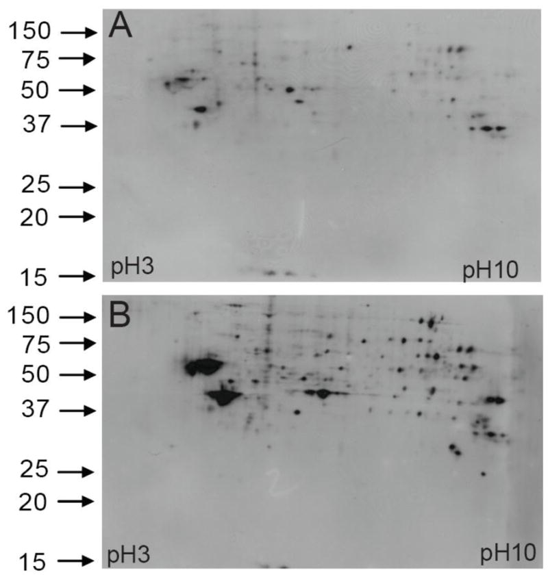
ICAT labeled protein pellets (100 μg) were first dissolved in a 2DE buffer and loaded onto IPG strips for isoelectric focusing (IEF). After IEF, the proteins on the IPG strips were reduced with DTT and alkylated with iodoacetamide, followed by the second dimension of separation in SDS gels. Subsequently, the proteins in each gel were transferred onto a nitrocellulose membrane. ICAT labeled SNO-proteins were detected by an anti-biotin antibody. A: Endogenous SNO-proteins isolated from SH-SY5Y cells. B: Increase of select SNO-proteins following SNO-Trx1 treatment, as seen from both the number and levels of SNO-protein signals detected.