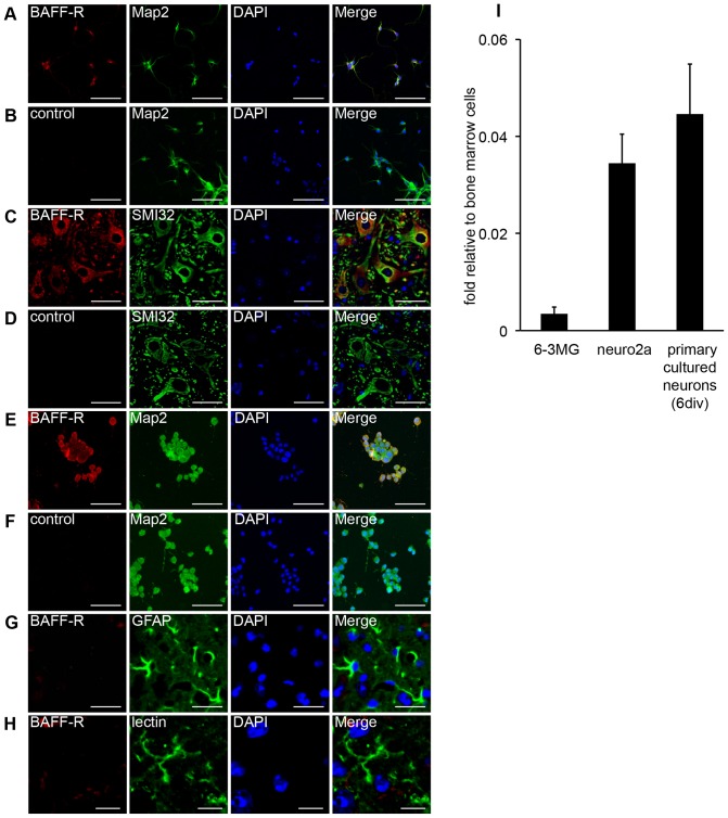
Figure 1. BAFF-R expression in mouse primary cultured neurons, on spinal cord neurons and on Neuro2a cells.
(A–F) Primary cultured mouse neurons (A and B), sections of a mouse spinal cord (C and D) and Neuro2a neuroblastoma cells (E and F) were co-stained with Cy5-conjugated anti–BAFF-R antibodies (A, C and E) or control antibodies (B, D and F) and an Alexa488-conjugated anti-microtubule-associated protein 2 (Map2) antibody (A, B, E and F) or anti-Neurofilament H Non-Phosphorylated (SMI-32) antibody (C and D). (G and H) Sections of a mouse spinal cord were co-stained with Cy5-conjugated anti–BAFF-R antibodies and an Alexa488 conjugated anti-GFAP antibody (G) or FITC-conjugated tomato lectin (H). 4′, 6-diamidino-2-phenylindole (DAPI) was also used to stain nuclei. Scale bars represent 100 μm (for panel A, B, E and F), 50 μm (for panel C and D), 25 μm (for panel G), and 10 μm (for panel H) respectively. (I) BAFF-R mRNA expression in 6–3 microglia cells, Neuro2a cells, and primary cultured neurons was examined by quantitative RT-PCR. The data are presented as the mean ± s.d. of samples examined in triplicate.

