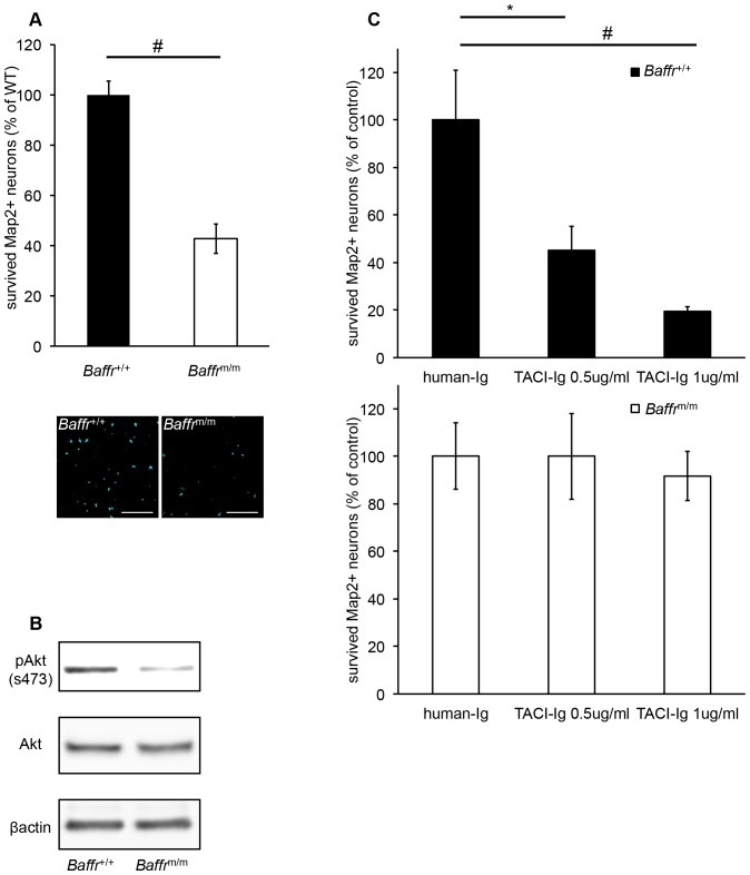
Figure 3. Role of BAFF-R in neuronal survival in vitro.
(A) The effect of a BAFF-R deficiency on neuronal survival. Neurons from E13.5 embryos of wild-type and A/WySnJ mice (Baffr m/m mice) were cultured for 7days under nutrient-limiting conditions, and then cell viability was measured by counting Map2+ viable neurons. Representative pictures are shown. Scale bar = 100 μm. (B) Reduced Akt phosphorylation in Baffr m/m neurons. After 7days of culture, wild-type and Baffr m/m neurons were assayed for the levels of total and phospho-Akt by immunoblot analysis. β-actin is shown as a loading control. (C) The effects of blocking BAFF-R on neuronal survival. Neurons from E13.5 embryos of wild-type and Baffr m/m mice were cultured with TACI-Ig or control human-Ig for 7days under nutrient-limiting conditions, and then Map2+ viable neurons were counted. Data are representative of three separate experiments. *:p<0.05, #:p<0.01.

