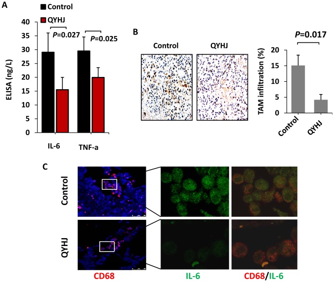
Figure 4. QYHJ suppresses cancer-related inflammation in pancreatic cancer.
A. An orthotopic nude mouse model was established and treated with QYHJ or vehicle as described above. Blood was collected from each mouse, and the serum concentrations of the pro-inflammatory cytokines IL-6 and TNF-α were detected by ELISA. The Student’s t-test was used to determine the statistical significance. B. To evaluate TAM infiltration, CD68 staining was performed using IHC. Original magnification, 200×. TAM infiltration was evaluated quantitatively by calculating the ratio of the CD68-positive area to the total area in each field, and the mean value from ten fields under 200× microscopy was determined. *P<0.05. C. Confocal microscopy of pancreatic cancer tissues treated with anti-CD68 (red) and anti-IL-6 (green). Cell nuclei were counterstained with DAPI.

