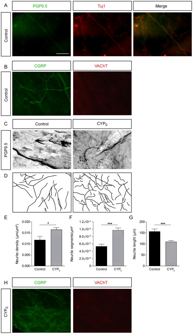
Figure 1. Sensory neurites sprouting in bladder wall in CYPc rats.
A, Nerve fibers stained for PGP9.5 and Tuj1 in whole mount urothelium - scale bar : 50 µm. B, Fibers are stained by CGRP antibody but not by VAChT antibody in control condition. C, PGP9.5 staining of whole mount bladder mucosa in control and CYP-treated rats. D, Line representation showing neurites based on the images in panel C, illustrating increased outgrowth in CYPc. E-G, Statistical comparison of neurite segments (E), neurite length (F) and neurite density (G) between control and CYPc rats. H, Neurite fibers observed in CYPc are stained by CGRP but by VAChT antibody.

