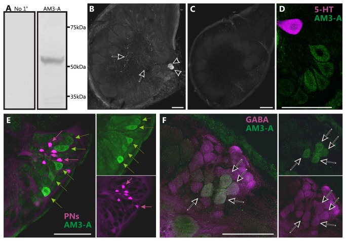
Figure 5. The Ms5HT1A receptor is expressed in a subset of GABA-like immunoreactive local interneurons.
(A) Western blots of the AM3-A antibody against Manduca protein and in which the AM3-A antibody was omitted (no 1o) produced no labeling. (B) Frontal section through an AL labeled using an antibody against a 5-HT1A receptor from prawn (AM3-A) generously provided by Dr. Maria Sosa. Note cell bodies (arrowheads; 24±2) and fine processes (arrows). (C) Manduca ALs labeled with AM3-A antibody pre-adsorbed with the Ms5HT1A receptor sequence (KDPDYLARITQQQKCLVSQD) homologous to the antigenic sequence used to generate the AM3-A antibody (KDPDFLVRVNEHKKCLVSQD) resulted in no labeling. (D) Double labeling against the Ms5HT1A receptor (green) and 5-HT (magenta) show no overlap. (E) Backfills of projection neurons (PNs) (magenta) reveal no co-localization of the Ms5HT1A receptor (green) in the lateral cell cluster of the AL. Magenta arrows indicate PNs and green arrows indicate Ms5HT1A-ir neurons. (F) Double labeling against the Ms5HT1A receptor (green) and GABA (magenta) revealed a subset of Ms5HT1A-ir cell bodies co-localizing GABA (arrows). All scale bars=100µm.

