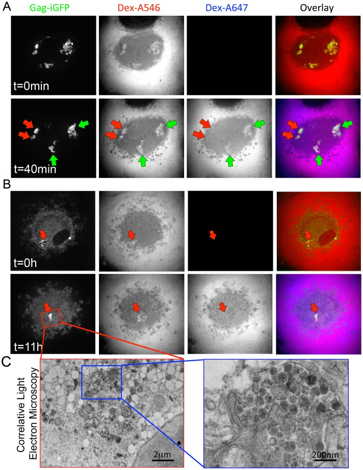
Figure 6. Transient connection of the Virus-Containing Compartment with the plasma membrane.
(A–C) Macrophages were infected for 4 days and imaged as in Fig. 3C. (A) Imaging was first performed after addition of a 10 kD Dex-A546 (t = 0 min, upper panel). A 10 kD Dex-A647 was added 40 min later, and imaging of the same cell was carried out (t = 40 min, lower panel). Several Gag-iGFP compartments are visible and appear accessible to both dextrans (green arrows) but others remain inaccessible to second dextran (red arrows). (B) Confocal imaging from 4 to 5 dpi, was performed immediately after addition of Dex-A546 (t = 0 min, upper panel). A Gag-iGFP+ sparse compartment containing Dex-A546 can be seen. After 11 hours (t = 11 h, lower panel), the same cell was exposed to Dex-A647 and imaged immediately after. The Gag-iGFP+ compartment appeared denser but remained negative for Dex-A647. (C) Correlative light and EM of the very same cell show that the GFP signal observed in light microscopy corresponds to a bona fide dense VCC containing viral particles.

