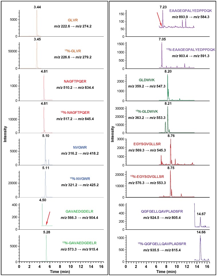
Figure 6. Ion-current profiles of mass transitions of eight tryptic peptides of hAPE1 and 15N-hAPE1 obtained using the tryptic hydrolysate of a protein fraction, which was collected during separation by HPLC of a nuclear extract of mouse liver.
The nuclear extract was spiked with an aliquot of 15N-hAPE1 prior to HPLC-separation. Peptides and monitored transitions are shown. The red arrows indicate the elution positions of GAVAEDGDELR and EAAGEGPALYEDPPDQK, which are absent, because they are not among the tryptic peptides of mAPE1.

