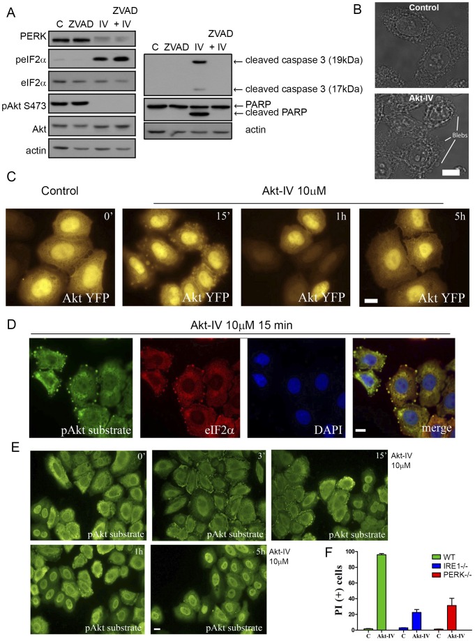
Figure 5. Akt-IV induces cell blebbing and a UPR dependent cell death.
(A) HEK293T cells were treated with DMSO (C), 100 µM of the caspase inhibitor ZVAD, 10 µM Akt-IV (IV) or both. PERK mobility, eIF2α phosphorylation, Akt phosphorylation on Ser473, caspase 3 cleavage and PARP cleavage were detected by WB. (B) Transmission images of HeLa cells treated for 15 min with DMSO (15 min) or with Akt-IV (10 µM), with blebs indicated. Bleb formation was clearly observed in HeLa and MEF cells but could not be detected in HEK293T cells. (C) YFP channel images of HeLa cells transfected with pAkt1-YFP plasmid and then treated for the indicated times with Akt-IV (10 µM). Akt1-YFP can be detected in blebs after 15 min of treatment. (D) HeLa cells were treated for 15 min with Akt-IV. Cells were fixed and immunostained against pAkt substrate/Alexa Fluor® 488 and total eIF2α/Alexa Fluor® 594. Green, pAkt substrate; Red, eIF2α; Blue, DNA. scale bar, 5 µm. (E) HeLa cells were treated for different times with Akt-IV (10 µM) and then cells were fixed and immunostained for pAkt substrate/Alexa Fluor® 488; scale bar, 5 µm. (F) MEF WT, IRE1−/− or PERK−/− were treated with Akt-IV (10 µM) for 12 h. Cell viability was measured by flow cytometry using propidium iodide. Data are representative of at least three independent experiments.

