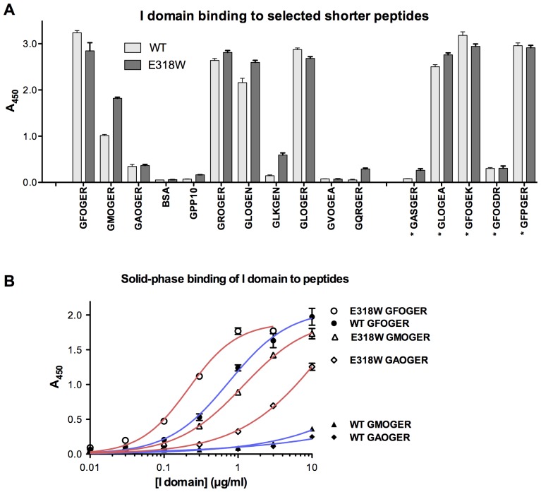
Figure 2. Binding of the integrin α2 I domain E318W mutant to selected peptides.
(A) Wild-type and E318W α2 I domains were used in binding assays as described in the legend to Fig. 1, with shorter triple-helical peptides as substrates. The sequence of the peptides is indicated on the x-axis, where an asterisk indicates sequences not found in collagens II and III. Six paired experiments were performed, each in triplicate, and data represent mean A450± SEM. (B) Increasing concentrations of wild-type and E318W I domains were applied to GFOGER, GMOGER and GAOGER coatings, and binding was measured as above. Curves shown are the best fit non-linear single-site binding curves, obtained using GraphPad Prism 5 for Mac, of 3 replicates in a single experiment.

