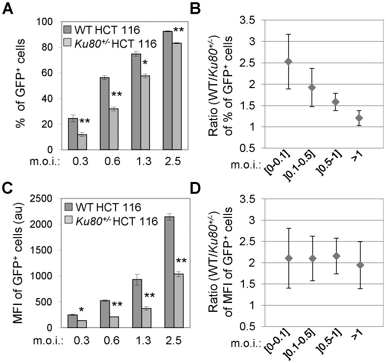
Figure 2. Ku80 haplodepletion reduces HIV-1-driven GFP expression.
(A–D) Wild-type (WT) and Ku80+/− human colon carcinoma HCT 116 cells were transduced at the depicted multiplicity of infection (m.o.i.) with XCD3 (HIV-1 env- nef - IRES-gfp) as indicated and were analyzed 48 h later by cytofluorometry for the percentage and the geometric mean fluorescence intensity (MFI) of green fluorescent protein-positive (GFP+) cells. Panels (A) and (C) present the results of the percentage and the MFI of GFP+ cells, respectively, obtained from at least two independent experimentations at the indicated m.o.i. (mean ± SD; *, p<0.05; **, p<0.01), except for the last m.o.i. for which one representative experiment (of 2 independent observations yielding similar results) is presented (mean ± SD). The ratios between WT and Ku80+/− HCT 116 cells for the percentage and the MFI of GFP+ cells at the depicted m.o.i. are reported in panels (B) and (D), respectively (mean ± SD, n = 3). au, arbitrary units.

