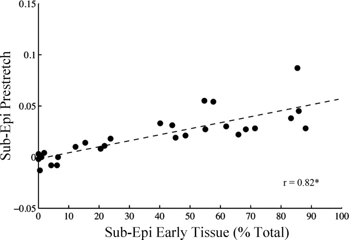
Fig. 4.
Fiber prestretch magnitude increases with the amount of earlier activated tissue at the subepicardium. Subepicardial peak fiber prestretch (Sub-Epi Prestretch) was correlated with the amount of subepicardial earlier-activated tissue (Sub-Epi Early Tissue), defined as percent total of the epicardial tissue area. Earlier activated tissue was determined from polar epicardial isochronal activation maps (see methods) in all animals during ventricular pacing at each site (i.e., 28 total data points). Linear regression analysis revealed a significant positive slope between Sub-Epi Prestretch and Sub-Epi Early Tissue. Sub-Epi, sub-epicardium (25% depth); r, correlation coefficient. *Slope different from zero (P < 0.001).

