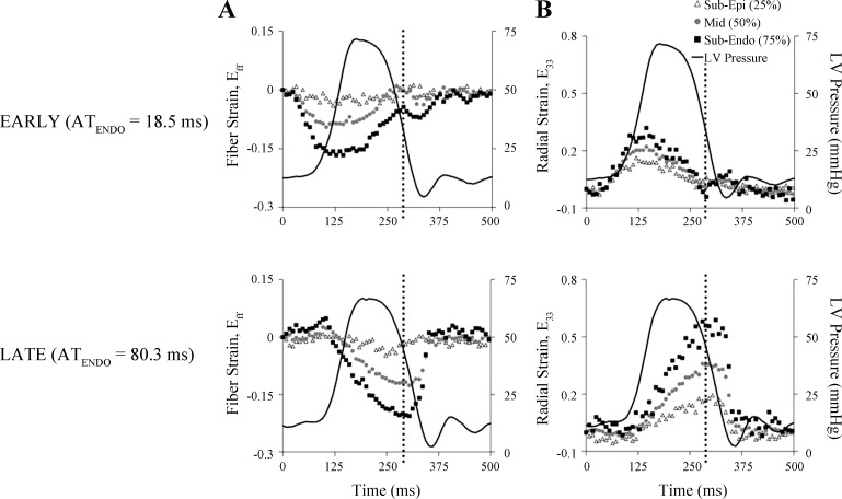
Fig. 5.
Differences in transmural fiber and radial strain time courses due to early and late electrical activation at the anterolateral left ventricle. Fiber strain (Eff; A) and radial strain (E33; B) at 3 transmural depths (subepicardium, midwall, and subendocardium) of the anterolateral LV are plotted throughout the cardiac cycle (in ms) for one representative animal (dog 9). Ventricular epicardial pacing at anterior apical (EARLY, top) and posterior basal (LATE, bottom) left ventricular sites resulted in marked differences in subendocardial activation time (ATENDO), as well as end-systolic strain values. Time of end systole is denoted by the black dotted line. LV pressure tracings are superimposed and synchronized with strain time courses. Reference time (0 ms) was the time of the local ventricular stimulus artifact. Sub-Epi, subepicardium (25% depth); Mid, midwall (50% depth); Sub-Endo, sub-epicardium (75% depth).

