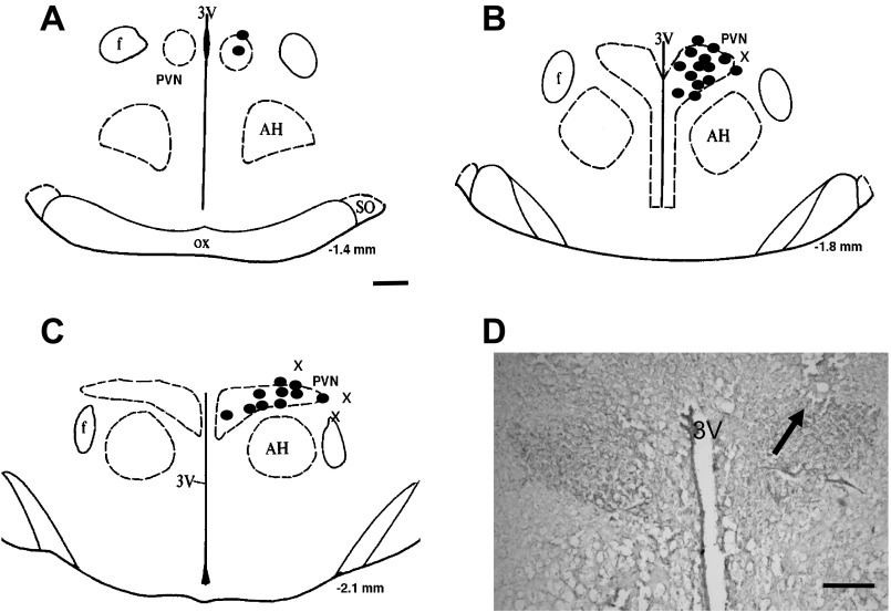
Fig. 6.
A–C: schematic representations of serial sections from the rostral (−1.4) to caudal (−2.1) extent of the region of the PVN. The distance (in mm) posterior to the bregma is shown for each section. Solid circles represent the site of termination of an injection that is considered to be within the PVN region. “x” represents the site of termination of an injection that is outside the PVN region. D: histological photo showing the injection site (arrow) in the PVN of one rat. AH, anterior hypothalamic nucleus; f, fornix; 3V, third ventricle; OX, optic tract; SO, supraoptic nucleus. Bar = 200 μm.

