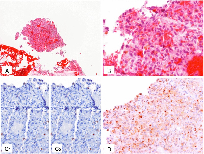
Figure 2.

Characteristics of Case 2. A, B: H&E of the metastatic cheek lesion, low (A) and high (B) power views; C: Negative IHC stains of S100 (C1) and Pan-Melanoma cocktail (C2) in the metastatic cheek lesion, scattered cells with non-specific staining present; D: Positive nuclear IHC staining of MITF in the metastatic cheek lesion.
