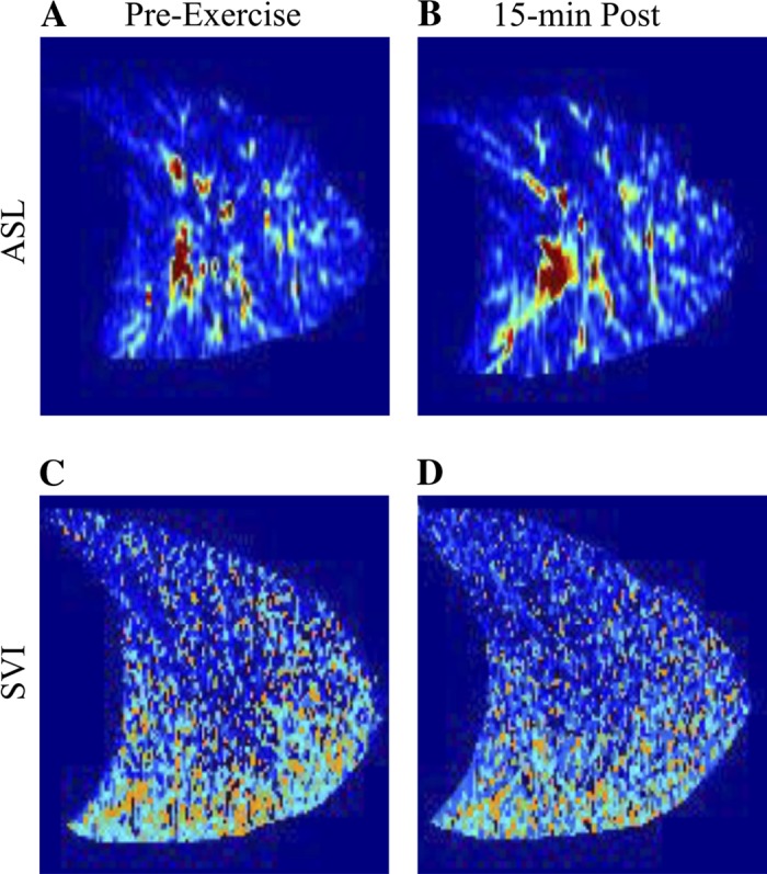
Fig. 2.

Representative supine images of pulmonary blood flow preexercise (A) and 15 min postexercise (B) and specific ventilation preexercise (C) and 15 min postexercise (D) in subject 6. In all images, the apex of the lung is to the right, and base of the lung is to the left. Signal from the large conduit blood vessels is removed from the blood flow images in postprocessing.
