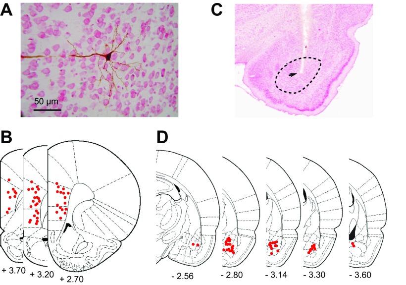
Fig. 3.
Example of a prefrontal cortex (PFC) neuron that was recorded from and filled with Neurobiotin. A: neutral, red-stained coronal section with a Neurobiotin-filled pyramidal neuron. B: overlay of 3 sections from The Rat Brain in Stereotaxic Coordinates, illustrating sites of recorded neurons (red dots). C: Nissl-stained section, illustrating a representative stimulating-electrode placement in the BLA (arrow). The black, dashed circle represents the boundaries of the BLA. D: overlay of sections of the The Rat Brain in Stereotaxic Coordinates, illustrating placements of BLA-stimulating electrodes (red dots). [From Paxinos and Watson (1998).]

