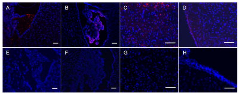
Fig 2. Brain distribution of Bacteriophage in brain.

Distribution of Clone 12-2 (A-D) and native M13 phage (E-H) in third ventricle (A,E), lateral ventricle (B,F), periventricular region of the third ventricle (C,G) and cortex (D,H) visualized 1h after i.v. administration in mouse caudal vein. Red: a secondary Cy3 conjugated Anti-mouse IgG; Blue: cell nuclei stained with 1μg/mL DAPI for 10min. The magnification bar represents 100μm.
