
Fig 3. Transmission electron micrographs of NP and TGN-NP.
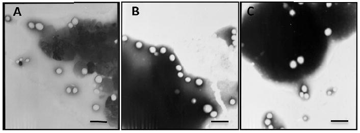
Different nanoparticles were negatively stained with phosphotungstic acid solution and observed under transmission electron micrographs: (A) NP; (B) TGN-NP (1:3); (C) TGN-NP (1:1). The bar is 200nm.