Fig. 4.

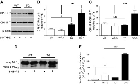
Phosphorylation of CPI-17 and 20-kDa myosin light chain (MLC20) is increased in mesenteric arteries of CPI-17-Tg mice. Mesenteric arteries were isolated from 12- to 14-wk-old male CPI-17-Tg and control WT mice. After carefully removing the surrounding fat, the mesenteric arteries were equilibrated in Krebs solution for 30 min. Next, the tissues were frozen at rest or after stimulation with 5-HT (10 μM) plus PE (100 μM) for 2 s as indicated. Proteins were separated by SDS-PAGE (A) or by glycerol gel electrophoresis (D). CPI-17 and MLC20 phosphorylation was determined by Western blot using antibodies against total CPI-17 (CPI-17-T), phospho-CPI-17 (CPI-17-P), or total-MLC20 (MLC20-T). A and D: representative Western blots. B and C are the quantification of CPI-17 phosphorylation normalized to β-actin or total CPI-17 protein, respectively. E: quantification of the percentage of the MLC20 that are phosphorylated under resting and after stimulation by 5-HT plus PE by using urea/glycerol gel electrophoresis; n = 4–10. S, stimulated with 5-HT plus PE; NS, not significant. *P < 0.05 and ***P < 0.001.
