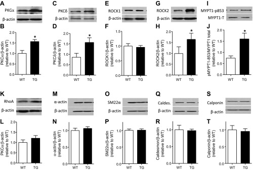
Fig. 5.

Protein kinase C (PKC) α/δ and rho kinase (ROCK) 2 proteins are increased in CPI-17-Tg mouse mesenteric arteries. Mesenteric arteries were isolated from 12- to 14-wk-old male CPI-17-Tg and control WT mice. After carefully removing the surrounding fat, the mesenteric arteries were homogenized, and the expression levels of PKCα/δ, rhoA, ROCK1/2 protein, pMYPT1-853, α-actin, SM22α, caldesmon, and calponin were determined by Western blot, respectively, shown in A, C, E, G, I, K, M, O, Q, and S. B, D, F, H, J, L, N, P, R, and T, respectively, are the quantifications of the blots of 3–11 independent experiments. *P < 0.05.
