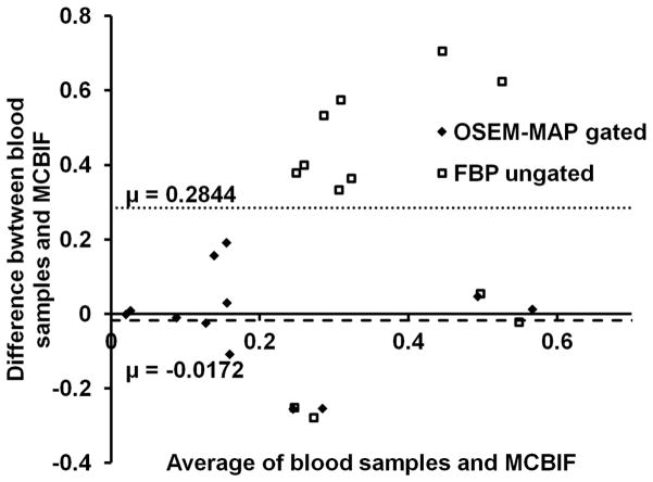
Figure 3. Residual plot.
MCBIF applied to high resolution gated images, when compared to the 2 late venous blood samples revealed an average difference of 0.017 MBq/cc in n=6 control mice. The precision (standard deviation of differences) was 0.13 MBq/cc. The figure also shows the analysis obtained from FBP un-gated images. The plot indicated an average difference and precision to be 0.28 MBq/cc and 0.33 MBq/cc respectively obtained from the FBP un-gated data sets. The residual plot indicates the quantitative accuracy and repetitive behavior of the MCBIF applied to OSEM-MAP gated images.

