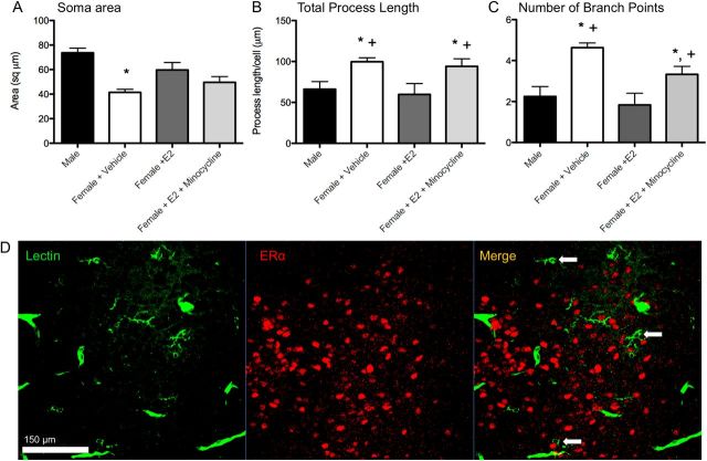
Figure 2.
Three dimensional morphometric analysis of microglia (A–C). Males and estradiol-treated females had significantly larger microglial cell bodies (A), shorter processes (B), and fewer process branch points (C) than vehicle-treated females in the POA on PN2. Cotreatment with estradiol and minocycline prevented estradiol's effects on process length and number of branch points. *Significantly different from males (ANOVA: p < 0.05). +Significantly different from females + E2 (ANOVA: p < 0.05). D, Microglia in the neonatal POA do not stain for estrogen receptor α. Confocal imaging at 40× magnification of POA sections costained for tomato lectin (a pan-macrophage stain) and estrogen receptor α in males (pictured) and females show no colocalization. Scale bar, 150 μm. Arrows indicate examples of labeled microglia that are negative for estrogen receptor α staining.

