Figure 2.

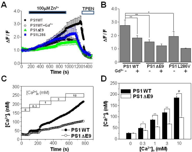
FAD-linked PS1 ΔE9 mutant attenuates Ca2+ influx mediated by Zn2+-permeable TRPM7. (A) Zn2+ influx was measured from HEK293 cells stably transfected with PS1 WT (n = 7), PS1 ΔE9 (n = 8), or PS1 L286V (n = 8). Also Zn2+ influx was measured from PS1 WT cells (n = 7) in the presence of 10 μM Gd3+. The intensity of Fluo Zin-3 gradually increased by the addition of 100 μM Zn2+ into the extracellular solution. The addition of membrane permeant chelator for Zn2+, TPEN, reduced the fluorescence intensity in all of cells tested. (B) The normalized fluorescence intensities were compared from different conditions as in (A). Zn2+ influx was also measured from PS1 ΔE9 (n = 7), or PS1 L286V (n = 7) cells in the presence of 10 μM Gd3+. (C) Cytoplasmic free Ca2+ concentration, [Ca2+]i, were compared between HEK293 cells transfected with either PS1 WT or PS1 ΔE9 using fura-2. Typical recordings are shown. Ca2+ influx were measured by adding back Ca2+ to the extracellular solution to obtain the indicated free Ca2+ concentrations. (D) When Ca2+ influx were compared they were larger in PS1 WT cells (n = 5) than in PS1 ΔE9 cells (n = 5). *, **, # represents p < 0.05, p < 0.01, p < 0.005 from paired t-tests, respectively.
