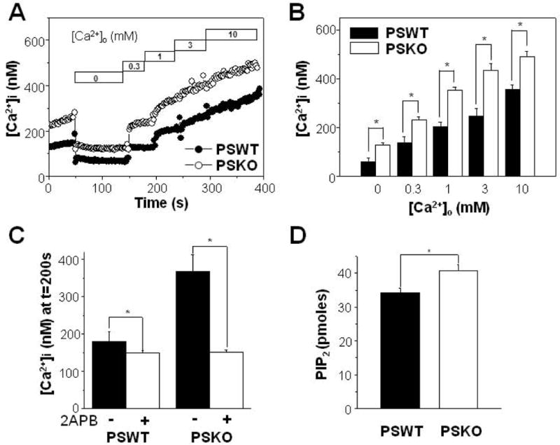
Figure 4.

Increased Ca2+ influx following the depletion of extracellular Ca2+ in PS1/2 double knockout (PSKO) MEF cells. (A) From this typical recording, PSKO cells showed increased resting [Ca2+]i either in the presence or in the absence of physiological [Ca2+]o compared to wild type PS1/2-transfected (PSWT) cells. Ca2+ influx were measured as in Fig. 2C by adding back Ca2+ to the extracellular solution as indicated. (B) Ca2+ influx were larger in PSKO MEF cells (n = 5) compared to PSWT cells (n = 5). Ca2+ influx was measured as in (A). (C) The effects of 2APB on Ca2+ influx were compared between PSWT (n = 8) and PSKO MEF cells (n = 8) as described in Fig. 3A. 2APB was able to block larger amount of Ca2+ influx from PSKO cells. (D) Steady state levels of PIP2 in membrane were assayed by using and ELISA kit from PSKO MEF cells (n = 4) and to PSWT MEF cells (n = 4). * represents p < 0.05 from paired t-test.
