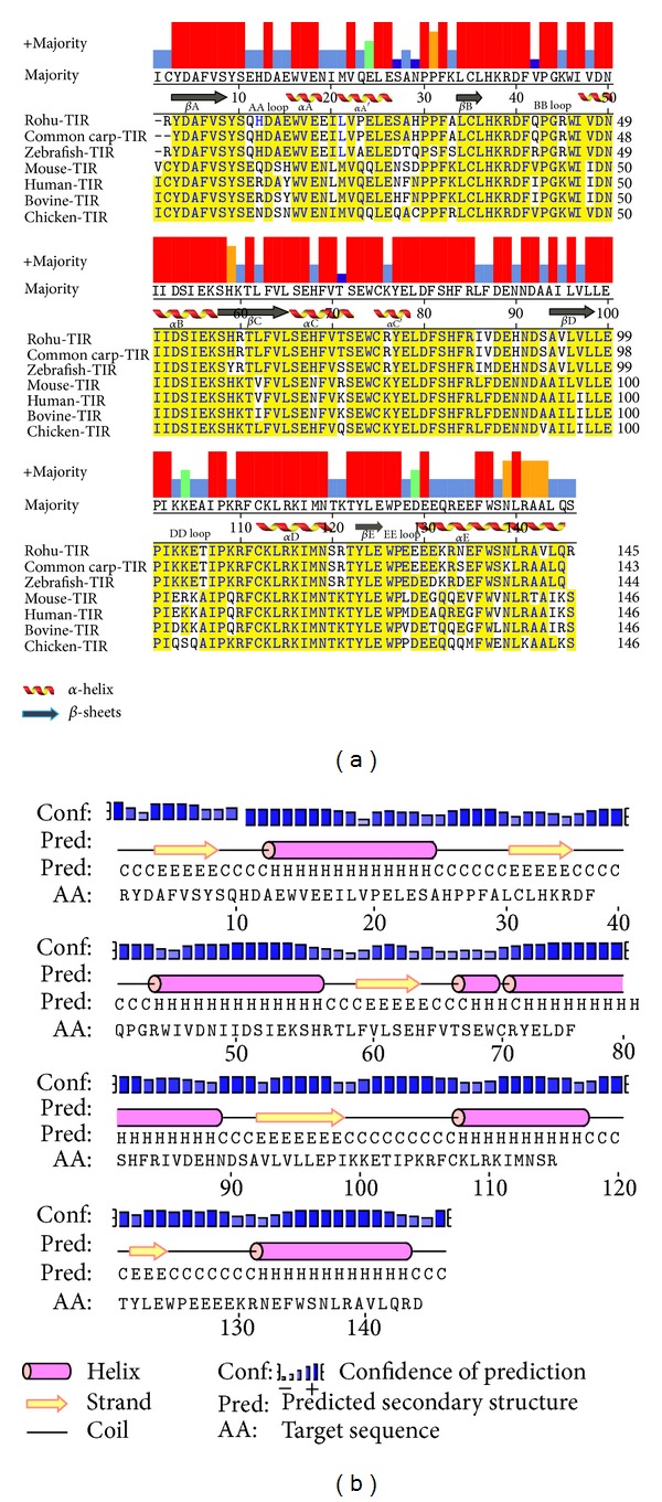
Figure 1.

Multiple sequence alignment and secondary structure prediction of TLR2-TIR domain. (a) Multiple sequence alignment of TLR2-TIR domain of rohu with others by MegAlign program. Conserved residues were shown in yellow. Consensus residues are shown in the majority axis. (b) Secondary structure representation of TLR2-TIR domain by PSIPRED. Helices denoted as “H,” beta strands as “E,” and loops as “C.”
