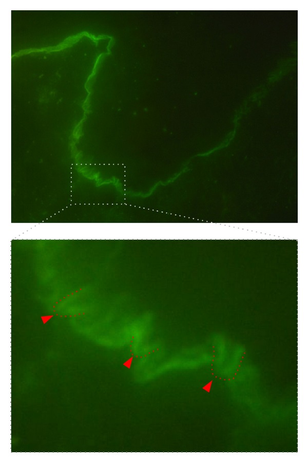
Figure 6.

u-serrated pattern in direct IF microscopy in EBA. Direct IF microscopy from perilesional EBA skin (staining for IgG, 400x original magnification). A linear binding along the dermal-epidermal junction is evident. The insert further magnifies the u-serrated binding of the autoantibodies (highlighted in red), which can also be observed in the original direct IF microscopy photograph.
