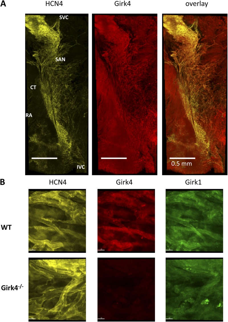
Figure 1.
Distribution and localization of anti-Girk immunoreactivity in isolated mouse atrio-sinus preparations. (A) Representative images of n = 3 isolated SAN right atrium (RA) preparations (endocardial side) double stained with HCN4 and Girk4 antibodies. CT, crista terminalis; IVC and SVC, inferior and superior vena cava, respectively. (B) Close-up views of pacemaker cells within the SAN stained with HCN4, Girk4, or Girk1 antibodies. Individual HCN4-positive cells within the SAN region of WT hearts showed strong membrane-associated Girk4 and Girk1 immunoreactivity (top). Girk4−/− SAN cells displayed HCN4-positive and Girk4-negative staining and no membrane-bound Girk1 (bottom). Bars, 10 µm.

