Figure 2.
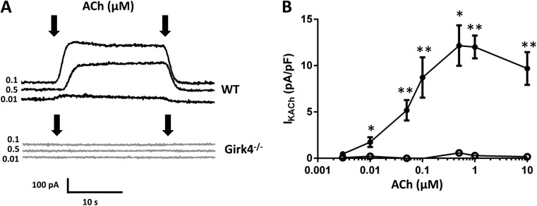
Inactivation of Girk4 channels ablates IKACh in mouse SAN cells. (A) Sample traces of IKACh current in WT and Girk4−/− SAN cells. IKACh was activated by ACh in WT and Girk4−/− SAN cells. Arrows indicate the period of time for which ACh was added. (B) IKACh density at different ACh concentrations in SAN cells from WT (0.003–10 µM; n = 4–16; closed circles) and Girk4−/− (0.003–10 µM; n = 3–13; open circles) mice. Error bars represent SEM. Statistical symbols: *, P < 0.05; and **, P < 0.01.

