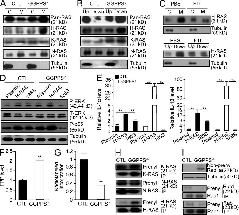
Figure 7.
GGPPS deletion results in increased H-Ras farnesylation in Sertoli cells, leading to the spontaneous hyperinflammatory response in the testes. (A and B) The membrane association and prenylation of Ras proteins in GGPPS−/− Sertoli cells were analyzed using membrane separation and Triton X-114 extraction methods, respectively. (C) The membrane association and prenylation of H-Ras in GGPPS−/− Sertoli cells after FTI treatment. (D) Erk1/2 and NF-κB activation (E) IL-1α (left to right: **, P = 0.000424; **, P = 0.0027; **P = 0.00249; **, P = 0.00227) and IL-1β (left to right: **, P = 0.00024; **, P = 0.0032; **, P = 0.000312; **, P = 0.0003) expression level. (F) The FPP level in Sertoli cells detected with LC-MS method (**, P = 0.000118). (G) Radiolabeled incorporation of FPP in GGPPS-deleted Sertoli cells (**, P = 0.00158). (H and I) IP analysis of RAS prenylation of Ras family (H) and Rap1a, Rac1, and Rabs. (I) The prenylation of N-Ras and K-Ras that can be either geranylgeranylated or farnesylated (H). The prenylation of Rap1a, Rac1, and Rabs that can only be geranylgeranylated (I). All of the groups of experimental animals contained a minimum of five mice. All of the data are representative of at least three replicates and are presented as the mean and SEM. *, P < 0.05, versus control; **, P < 0.005, versus control.

