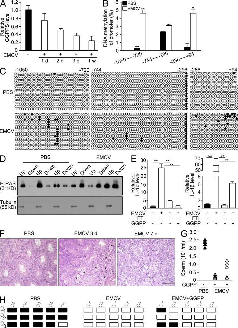
Figure 9.
EMCV challenge results in a mouse testis defect due to altered protein prenylation by inhibiting GGPPS expression. (A) GGPPS expression in mouse testes after EMCV challenge over time. (B) The methylation level of CpG islands in GGPPS’s promoter after EMCV infection (**, P = 0.00487; *, P = 0.0136). (C) The prenylation of H-Ras in Sertoli cells after EMCV infection. (D) The expression of IL-1α (PBS versus EMCV, **, P = 0.000339; EMCV versus EMCV+FTI, **, P = 0.000543, EMCV versus EMCV+GGPP, **, P = 0.000344) and IL-1β (PBS versus EMCV, **, P = 0.00277; EMCV versus EMCV+FTI, **, P = 0.00273; EMCV versus EMCV+GGPP, **, P = 0.003675) after EMCV infection after GGPP or FTI administration. (E) H&E staining after EMCV infection. Stars indicate the degenerated tubules. (F and G) Fertility after GGPP supplements into EMCV-challenged mice. Sperm number after GGPP administration (F) and pregnancy of mice after GGPP administration into EMCV-infected mice (G). (H) Pregnancy analysis of ECMV-attacked mice after GGPP administration. All of the groups of experimental animals contained a minimum of five mice. All of the data are representative of at least three replicates and are presented as the mean and SEM. *, P < 0.05, versus control; **, P < 0.005, versus control. Bar, 100 µm.

