
Figure 3. Mechanisms of EPEC induced Inhibition of SERT.
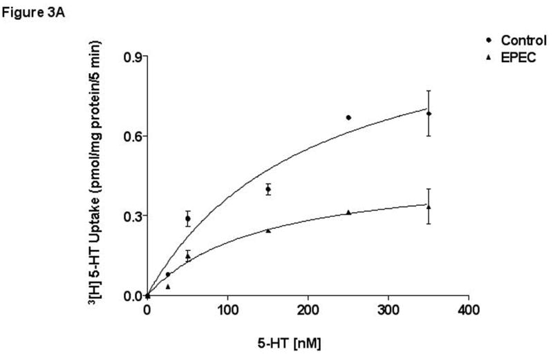
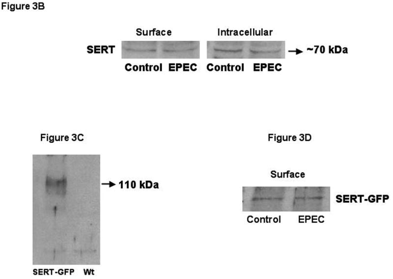
A)Kinetic studies of EPEC induced inhibition. A Michaelis–Menten plot from a representative experiment is shown. N=5. * P < 0.01 compared to control. B) Cell-surface expression of SERT. Caco-2 monolayers were infected with wild-type EPEC for 60 minutes and subjected to biotinylation. C) SERT expression in transiently transfected Caco-2 cells. SERT expression in wild type untransfected and cells transiently transfected with SERT-GFP construct utilizing ant-GFP antibodies. D) EPEC does not alter cell-surface expression of SERT-GFP: Representative blots from 3 different experiments are shown.