
Figure 6. SERT immunostaining in EPEC infected mouse small intestine.
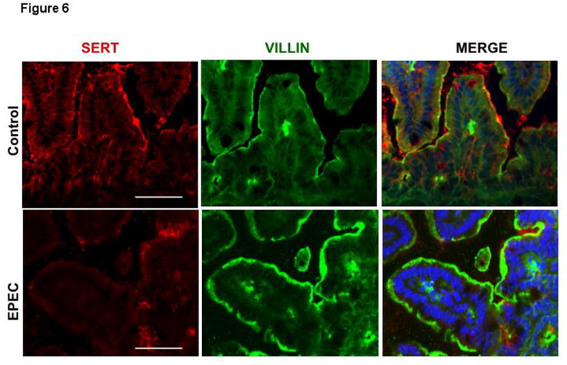
Immunofluorescent staining for SERT (red) and villin (green) with blue counterstained nuclei was performed on OCT sections of control and EPEC infected mouse distal small intestine. Scale bar = 50 μm. A representative of 4 different experiments is shown.