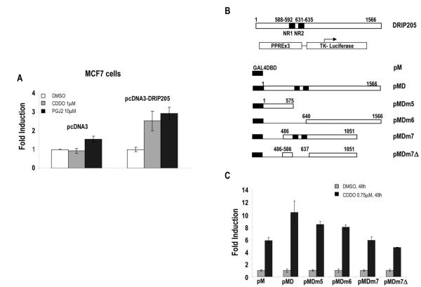
Fig. 4.
A. MCF-7 cells were transiently co-transfected with 1μg of pPPRE-TK-LUC and 4μg of pcDNA3 or pcDNA3-DRIP205, treated with indicated concentrations of CDDO or 15dPGJ2 for 92.5 hours, after which PPARγ transactivation was determined by relative luciferase activity calculated by dividing luciferase activity by protein concentration for each well. Results are shown as mean ± SEM for three separate experiments. B. Map of GAL4-DRIP fusion proteins. C. Coactivation of PPARγ by GAL4-DRIP fusion proteins. SW480 cells were co-transfected with 1000 ng of pPPRE3-LUC, 250 ng of β-galactosidase, 500 ng of pM empty or pM DRIP deletion mutants, treated with DMSO and 0.75μM CDDO, and luciferase activity was determined. Results are shown as mean ± SEM for three separate experiments.

