Fig. 5.
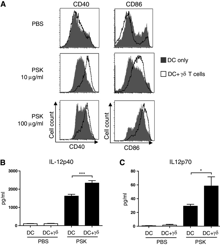
Reciprocal activation of DC in the BMDC-γδ T cell co-culture leads to increased expression of co-stimulatory molecules and enhanced production of IL-12. BMDC and BMDC-γδ T cell co-culture were treated with PSK for 24 h, and the cells were harvested for FACS analysis of DC activation markers. The culture supernatant was analyzed for IL-12 using ELISA. a Expression of CD40 and CD86 in BMDC treated with control RPMI medium or PSK (10 or 100 μg/ml). Shaded histograms: DC alone culture; empty histograms: DC + γδ T cell co-culture. b, c The levels of IL-12p40 and IL-12p70 in control untreated DC or DC + γδ T cell co-culture (white columns) and PSK-treated DC or DC + γδ T cell co-culture (black columns). *p < 0.05, by two-tailed Student’s t test

