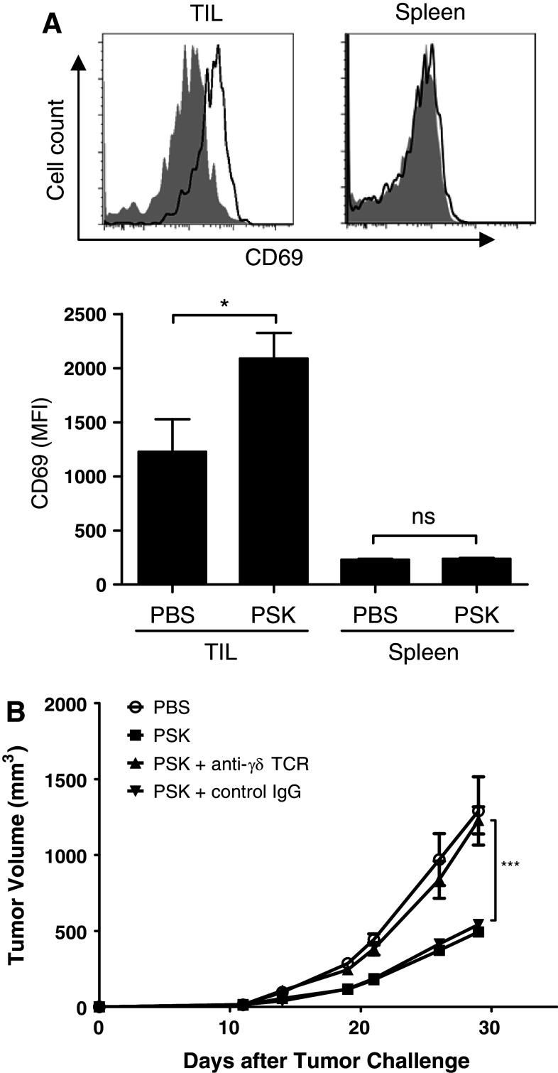
Fig. 7.
In vivo PSK treatment activates γδ T cells in TIL, and γδ T cell contributes to the anti-tumor effect of PSK. a Intratumoral injection of PSK results in CD69 upregulation on γδ T cells in TIL, but not in spleen. Shown are representative overlay histograms of CD69 expression on γδ T cells in TIL or spleen from control PBS- and PSK-treated mice. Shaded histogram PBS-treated mice (control); empty histogram: PSK-treated mice. The summary bar graph shows the mean fluorescence intensity (MFI) of CD69 expression in γδ T cells (mean ± sem) in TIL and spleen in PBS or PSK group (n = 8 mice per group). *p < 0.05, by two-tailed Student’s t test. b Depletion of γδ T cells during PSK treatment decreased the anti-tumor effect of PSK. Mice received anti-γδ TCR mAb or a control hamster IgG during PSK treatment. PSK by itself significantly inhibited tumor growth, and the effect is significantly attenuated when mice received anti-γδ TCR mAb. **p < 0.01 between PSK + γδ T cell depletion group (filled triangle) and PSK group (open square); ***p < 0.0001 between control PBS (open circle) and PSK group (open square). There was no difference between PSK + control IgG (filled inverted triangle) and PSK group (open square). N = 5 mice per group. Similar results were obtained from two independent experiments

