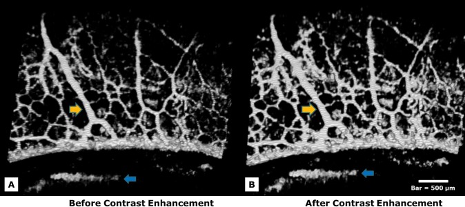
Figure 2. .
Several straight radial arcades draining vessels perpendicular to the limbal margin are shown (yellow arrows). Contrast enhancement was applied to SD-OCT images to increase image clarity. Enhancing contrast significantly increased both the contrast associated with vessels (yellow arrows) and also the contrast of noise (blue arrows). (A) An image is shown prior to contrast enhancement. (B) The same image is shown after contrast enhancement. Manual contrast adjustment followed in both SD-OCT and fluorescence images until an optimal balance was achieved. Bar = 500 μm.

