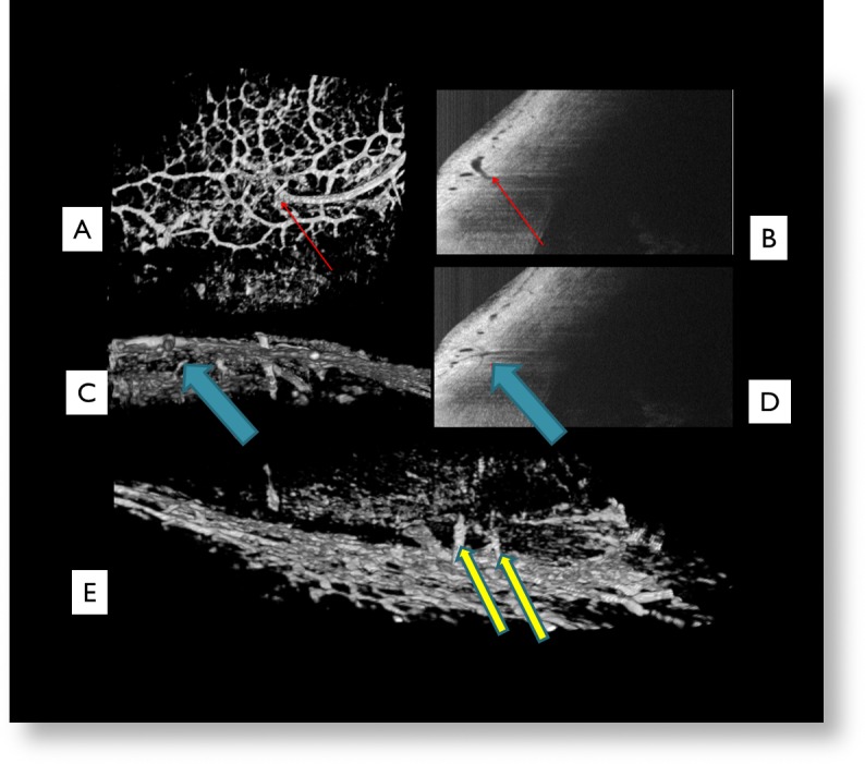
Figure 3. .

The superficial venous plexus visualized was composed of a series of small interconnected venules between 25 and 100 μm in diameter with many interconnecting branch points forming a dense vascular hexagonal meshwork (A). Red arrows indicate a vessel seen on the virtual casting (A) and its corresponding location in B-scan (B). Blue arrows indicate a suspected aqueous vein (C) descending from the superficial ISVP to the midlimbal ISVP and its corresponding location in B-scan (D). Yellow arrows indicate two suspected aqueous veins seen in this 180 degree rotated virtual casting image (E).
