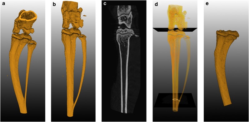
Figure 4.
Selecting a VOI using orthogonal cross-sections. (a) 3D rendering of the original scan volume. (b) 3D rendering of the complete image stack of cross-sections generated perpendicular along the centerline. (c) Longitudinal cross-section generated from the reformatted space. The sectioning plane can be rotated around the longitudinal axis. These sections can be used for side-by-side comparison between different scans. (d) Definition of transversal cutoff planes using the reformatted space to calculate the relative position between the knee and the branch point of the fibula and tibia. (e) 3D rendering of a VOI. The volume was selected in the original space using a region grower limited by the cut off planes, which were mapped back from the reformatted space. Adapted with permission from Snoeks et al.16

