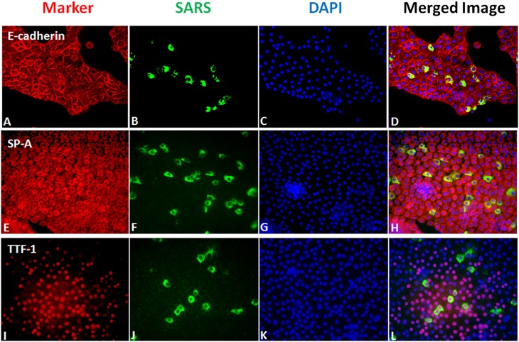
Figure 2.
Immunofluorescent staining for SARS-CoV nucleocapsid protein and selected alveolar type II cell markers. Cells were grown under A/L conditions as described in Materials and Methods, inoculated with SARS-CoV at an estimated multiplicity of infection of 2, and fixed 24 hours after inoculation. (A–D) Staining for E-cadherin (A), SARS-CoV (B), DAPI (C), and merged (D). (E–H) Staining for SP-A (E), SARS-CoV (F), DAPI (G), and merged (H). (I–L) Staining for TTF-1 (I), SARS-CoV (J), DAPI (K), and merged (L). Cells that are infected with SARS-CoV stain for type II cell markers.

