
Figure 7.
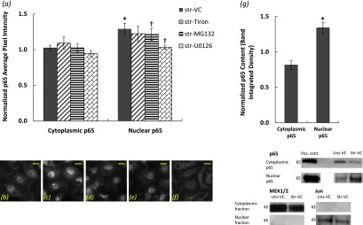
(a) Quantification of cytoplasmic and nuclear p65 subunit of NF-κB content in type I–like RAECs that were stretched at 37% ΔSA for 60 minutes and treated with VC (n = 19), the superoxide scavenger tiron (n = 7), the NF-κB activation inhibitor MG132 (n = 14), and the ERK inhibitor U0126 (n = 3) based on an image-processing algorithm applied on the p65-DAPI images. (b–f) Representative images of the p65 subunit of NF-κB in type I–like RAECs that remained unstretched and treated with VC (b). (c–f) Cells that were stretched for 60 minutes at 37% ΔSA and treated with VC (c), tiron (d), MG132 (e), and U0126 (f). Bar = 10 μm. (g) Quantification of cytoplasmic and nuclear p65 subunit of NF-κB content in type I–like RAECs that were stretched at 37% ΔSA for 60 minutes based on Western blotting (see representative bands below the graph). Cytoplasmic p65 was normalized to the cytoplasmic marker protein MEK1/2, and nuclear p65 was normalized to the nuclear marker protein Jun. Data are expressed as mean ± SEM of fold change above unstretched cells treated with VC. *P < 0.05 versus unstretched VC; †P < 0.05 versus stretched VC.